JCRB0138 KHM-3S
Cell information
Cell type:general cells (View Pricing Information)
| JCRB No. | JCRB0138 | Cell Name | KHM-3S |
|---|---|---|---|
| Profile | small cell carcinoma, HTLV-1gene integration | Other Name | |
| Animal | human | Strain | |
| Genus | Homo | Species | sapiens |
| Sex | M | Age | 58 |
| Identity | available | Tissue for Primary Cancer | lung (cancer) |
| Case history | small cell lung cancer whose serum contained high levels of soluble interleukin-2 receptors. | Metastasis | |
| Tissue Metastasized | Genetics | No OKT11,OKT4,OKT8,B1,& B4 found. NKH-1, rearranged T cell receptor and HTLV-1 integration observed. | |
| Life Span | infinite | Crisis PDL | NT |
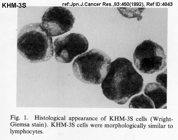
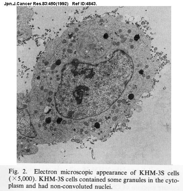
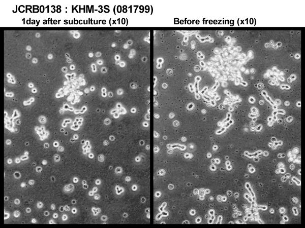
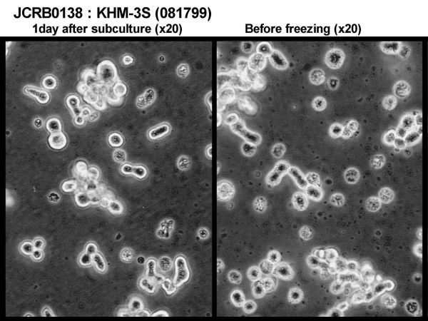
| Morphology | lymphocyte-like | Character | HTLV-1 positive small cell lung cancer cells. |
| Classify | tumor | Established by | Matsuzaki,H. |
| Registered by | Matsuzaki,H. | Regulation for Distribution | HTLV-1 positive |
| Comment | Year | 1999 | |
| Medium | RPMI1640 medium with 20 % fetal calf serum was used at the begining. The FCS concentration was reduced to 10% when the cells began to grow steadily. | Methods for Passages | Simple dilution. Suspension culture. |
| Cell Number on Passage | 1-2 x 10^5 cells/ml | Race | |
| CO2 Conc. | 5 % | Tissue Sampling | pleural effusion |
| Tissue Type |
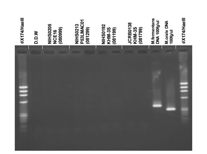
| Detection of virus genome fragment by Real-time PCR | |||||||||
|---|---|---|---|---|---|---|---|---|---|
| Detected DNA Virus | tested | Detected RNA Virus | tested | ||||||
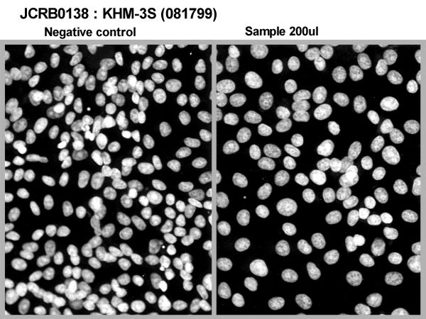
| CMV | - | parvoB19 | - | HCV | - | HTLV-1 | + | ||
| EBV | - | HBV | - | HIV-1 | - | HTLV-2 | - | ||
| HHV6 | - | HTLV-1 | + | HIV-2 | - | HAV | - | ||
| HHV7 | - | HTLV-2 | - |
-/negative. +/positive. nt/not tested. (positive (+) does not immediately mean the production of infectious viral particles.) |
|||||
| BKV | - | HIV-1 | - | ||||||
| JCV | - | HIV-2 | - | ||||||
| ADV | - | HPV18 | - | ||||||
| Notes | The integration of HTLV-1 proviral DNA is observed in KHM-3S, but the HTLV-1 envelope protein is not detected in the surface of KHM-3S (Jpn J Cancer Res. 1992 ay;83(5):450-457, PubMed ID 1319985). | ||||||||
| Reference | |
|---|---|
| Pubmed id:1319985 | Human T-cell leukemia virus-1-positive cell line established from a patient with small cell lung cancer. Matsuzaki H,Hata H,Asou N,Yoshida M,Matsuno F,Takeya M,Yamaguchi K,Sanada I,Takatsuki K Jpn J Cancer Res. 1992 May;83(5):450-7 |
| Pubmed id:2169997 | Human T-cell leukemia virus type 1 associated with small cell lung cancer. Matsuzaki H,Asou N,Kawaguchi Y,Hata H,Yoshinaga T,Kinuwaki E,Ishii T,Yamaguchi K,Takatsuki K Cancer. 1990 Oct 15;66(8):1763-8 |
| Images |
|---|
              |
LOT Information
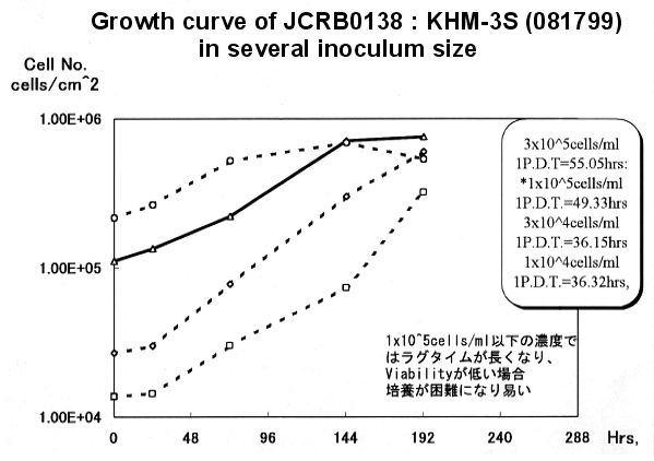
Viability/Growth rate/Cell number are represented as actual values measured at lot presentation in JCRB, but are not guaranteed values. Additionally, the doubling time is a rough value measured during passages.| Cell No. | JCRB0138 | Cell Name | |
|---|---|---|---|
| LOT No. | 03052003 | Lot Specification | distribution |
| Medium | RPMI1640 medium with 10% fetal bovine serum (FBS lot; BioWhittaker 0S069F) | Temperature | 37 C |
| Cell Density at Seeding | 1.0 x 10^5 cells/ml | Methods for Passages | Dilution, passage by 4 days interval. |
| Doubling Time | D.T. = ca. 2days | Cell Number in Vial (cells/1ml) | 7.9 x 10^5 |
| Viability at cell freezing (%) | 66.5 | Antibiotics Used | free |
| Passage Number | Unknown (11 at bank) | PDL | |
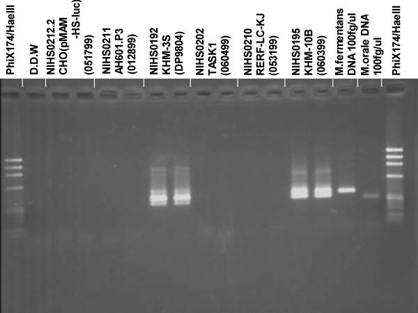
| Sterility: MYCOPLASMA | - | Sterility: BACTERIA | - |
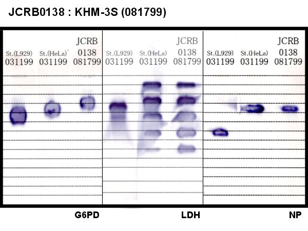
| Sterility: FUNGI | - | Isozyme Analysis | Confirmed as human by NP, G6PD (type B), LD. |
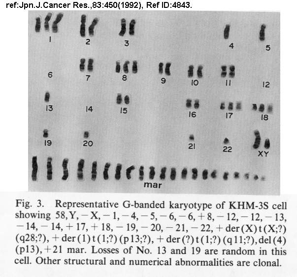
| Chromosome Mode | Chromosome Information | ||
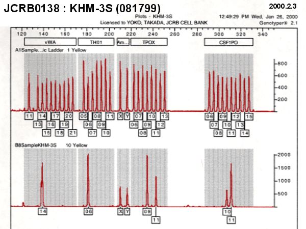
| Surface Antigen | DNA Profile (STR) | ||
| Adhesion | No | Exoteric Gene | |
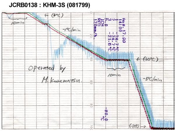
| Medium for Freezing | 10% DMSO, 20% FBS-culture medium | CO2 Conc. | 5% |
| Viability immediately after thawing (%) | Additional information |